Research Article 
 Creative Commons, CC-BY
Doppler Flow Velocity Measurements in the Extracranial Internal Carotid and Vertebral Arteries in Healthy Adults
*Corresponding author: Sanja Kovacic, Department of Neurology, General Hospital Zabok and Hospital of Croatian Veterans, Bracak 8, 49210 Zabok, Croatia
Received: September 21, 2022; Published: September 28, 2022
DOI: 10.34297/AJBSR.2022.17.002322
Abstract
Objective: To analyze normal hemodynamics in the extracranial part of the internal carotid arteries (ICA) and vertebral arteries
(VA) with reference to age and gender.
Methods: Angle-corrected flow velocities (peak-systolic velocity, PSV and end-diastolic velocity, EDV) and VA diameter were
measured, and waveform parameters calculated (pulsatility index, PI and resistive index, RI) on 227 healthy adults (125 women).
Results: Except for a slight increase of PSV in the left VA in men, and wider left VA diameter in both genders, there were no
clinically significant side-to-side differences in the investigated parameters. In 10-year periods, expected decreases in PSV ICA, EDV
ICA, PSV VA, and EDV VA were 3-3.7, 2.2-2.8, 0.6-1.5, and 0.7-1 cm/s, respectively while expected increases in RI ICA, PI ICA, RI VA,
PI VA, and VA diameter were small, in a range of 0.01-0.07. Average velocities were higher in women up to 60 years on both sides
in ICA and VA. In the women, expected values of PSV ICA, EDV ICA, PSV VA, and EDV VA were 5.9-6.2, 1.9-2.2, 2.1-4.3, and 1 cm/s,
respectively, i.e., higher than in the men. PI and RI showed no association with gender. Measurements showed that PSV ICA, EDV ICA,
PSV VA, and EDV VA ranged 40-108, 10-39, 24-63, 2-22 cm/s in women and 35-114, 7-36, 24-66, and 5-18 cm/s in men, respectively.
RI ICA, PI ICA, RI VA, PI VA, and VA diameter were in the ranges 0.5-0.8, 0.9-1.9, 0.6-21, 1.1-24, and 1.4-4.9 in women; 0.5-0.9, 0.8-2.1,
0.6-0.9, 1.1-2 mm, and 2.1-5.3 mm in men, respectively.
Conclusion: Velocity was higher in the women up to 60 years. In older age groups the flow rates equalized in both genders. EDV
ICA decreased with age in both genders. PSV ICA showed a declining trend only in women. RIs and PIs showed a slight tendency to
increase with age in both genders. In the vertebral arteries, a slight tendency to decreased flow velocities with age was seen mostly
in EDV in both genders, whereas PSV showed decline and diameter increase only in women.
Keywords: Hemodynamics; Internal carotid arteries; Flow velocities
Introduction
Carotid Doppler ultrasonography is a powerful modality for evaluating the pathology of carotid and vertebral arteries. Mainly, it represents a good tool for visualizing the blood flow in the vessel and detecting stenotic segments. A vast range of hemodynamic data can be generated by Doppler ultrasound. Adjusting the velocity range is one of the important ways of controlling Doppler ultrasonography parameters. The Doppler spectrum supplies several functions, maximum velocity values as peak systolic velocity (PSV) and enddiastolic velocity (EDV), which refers to the highest time-averaged mean velocity over the complete cardiac cycle envelope [1]. The peak systolic velocity threshold is considered the most significant factor in the diagnosis of vascular stenosis diseases [2].
Except for the oxygen consumption and peripheral vascular resistance of the distal microcirculation, cardiac function, blood pressure, anxiety and medications, blood flow in the vessel is influenced by the vessel wall compliance (resistive index, RI) exerted by the vessel wall and the pressure variation (pulsatility index, PI), which issued to express the relationship between blood flow pulsation and arterial pulsation [3,4]. The PI and RI calculation methods are not affected by the Doppler flow angle. The PI and RI can be applied to represent both the elasticity and resistance of downstream and upstream blood vessels.
Some previous studies proved the presence of age- and genderdependent changes in the PSV and EDV in the extracranial carotid and vertebral arteries [5-7].
Although much data has been published, there is still insufficient research on normal carotid and vertebral blood flow velocities and indices, while the decline of the velocity with age is still a subject of controversy [7,8]. Therefore, there is a need to record normal cerebral blood flow parameters in healthy adults in the context of the effects of age and gender. Furthermore, in patients with cerebrovascular diseases, it is worth knowing the normal range all the parameters included in the calculation of carotid and vertebral arterial pulsatility and resistance, not only the most frequently researched the PSV values, but also the EDV values. Resistive index (RI) and pulsatility index (PI) that were extracted from flow velocities are important hemodynamic parameters that have been simply and accurately used in the prognosis of cardiovascular complications [9]. The aim of this study was to investigate the natural effect of age and gender on flow velocities and waveform parameters in the extracranial arteries and luminal diameters of the vertebral arteries, as well as to provide more reference data for these parameters in different age and gender groups.
Materials and Methods
Ethics approval and consent to participate
The study was carried out in accordance with the Declaration of Helsinki. It was approved by the Medical Ethics Committee of the General Hospital Zabok and Hospital of Croatian Veterans, Zabok, Croatia. Informed consent to the procedure was obtained from each patient.
Study design, setting, and participants
The study was designed as a descriptive, cross-sectional, observational study. The study population involved 227 (102 men and 125 women) healthy adult volunteers aged 30-86, recruited from the Nursing School, the hospital staff, and their relatives. Exclusion criteria were all known risk factors for cerebrovascular disease (except for age, well-regulated arterial hypertension, and dyslipidemia), clinical and Doppler evidence of moderate or severe carotid atherosclerosis, abnormal hormonal and ECG status, abnormal echocardiography, anemia, fibromuscular dysplasia, pregnancy in female subjects, giant cell arteritis, and malignancies. The inclusion criteria for subjects older than 65 years was normal cardiac ultrasound.
Carotid duplex ultrasound scans were performed at the Vascular Laboratory of the General Hospital Zabok and Hospital of Croatian Veterans.
The extracranial arteries were explored using a 7.0-MHz linear-array transducer of a computed sonography system (AlokaProsoundAlpha6, Tokyo, Japan). The examiner was in a lateral sitting position and used his right hand for both carotid arteries. The patients were positioned in the optimal head position, at an angle of approximately 45° to the artery being examined. Both the B-mode and pulsed-signal wave Doppler (spectral Doppler) were utilized to measure the exact flow velocities. To obtain proper velocity measurements, an adequate acoustic angle of 45-60°was ensured. The VA diameter and center-line velocity waveform of the ICA and VA were recorded. We analyzed ICA velocity measurement data in the distal Doppler sample locations. In the vertebral arteries, the cross-sectional mean diameter and velocities were measured in the second part of the vertebral artery (cervical vertebrae levelsC4 to C5). All measurements were made with the vessel imaged in the longitudinal section with the maximum possible length shown, and with both anterior and posterior vessel walls visualized. PSV and EDV and the vertebral luminal diameter of the ICA and VA were measured manually, in accordance with the recommendations of some authors [10,11]. RI and PI were calculated according to standard equations [12-14]. All patients were examined by the same ultra-sonographer with 20 years of experience to avoid interobserver variability.
Statistical methods
After analyses of the distribution normality of the analyzed parameters, their distributions were presented as mean and standard deviations. Analyses of side-to-side differences for each sonographic parameter were performed using Student’s paired t- test. Associations of the PSV, EDV, PI, and RI of the ICA and AV as well as the diameter of the VA with age and sex were analyzed in a series of linear regression models. After the interpretation of regression coefficients, range values for the analyzed parameters were presented as median, interquartile range, minimum, and maximum for 10-year periods, separately for the women and men. For statistical data analyses, we used the statistical package Statistica 13.5 (StatSoft, TIBCO).
Results
Baseline characteristics
A total of 227 healthy adults were included in the study, 102 (44.9%) males and 125 (55.1%) females. The youngest participant was 30 and the oldest 86 years old.
Table 1 represents a side-to-side comparison of the average values of PSV, EDV, PI, and RI of the ICA and VA, and the luminal diameters of the VA. The values were normally distributed. The results showed no clinically significant side-to-side differences in the investigated parameters on both extracranial arteries, except for a very slight increase of PSV in the left VA in men, and wider left VA diameter in both genders. Although those differences were statistically significant, the effect sizes were small (effect size of the PSV VA = 0.3, effect size of the AV diameter in the females =0.26, effect size of the AV diameter in the males= 0.35).
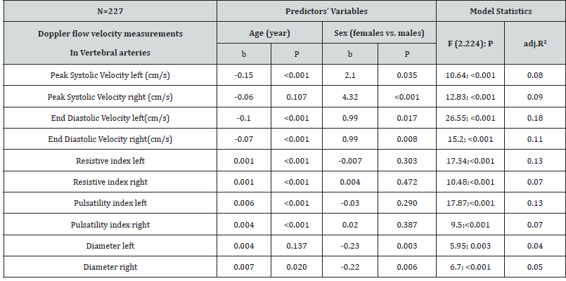
Regression analyses showed age-dependency of all variables on both sides in the ICA and VA, in terms of PSV and EDV decrease and a very slight RI and PI increase, except for PSV of the right VA and luminal diameter of the left VA. In 10-year periods, expected decreases in PSV ICA, EDV ICA, PSV VA, and EDV VA were 3-3.7, 2.2- 2.8, 0.6-1.5, and 0.7-1 cm/s, respectively. Expected increases in RI ICA, PI ICA, RI VA, PI VA, and VA diameter in 10-year periods were small, in a range of 0.01-0.07.
Average velocities were higher in women on both sides in ICA and VA. In the women, expected values of PSV ICA, EDV ICA, PSV VA, and EDV VA were 5.9-6.2, 1.9-2.2, 2.1-4.3, and 1 cm/s, respectively, i.e., higher than in the men. PI and RI showed no association with gender (Tables 2 & 3).
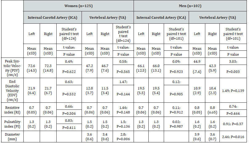
Table 1. Distribution and side-to-side comparison of PSV, EDV, PI, RI ICA and VA and luminal diameter of the AV.
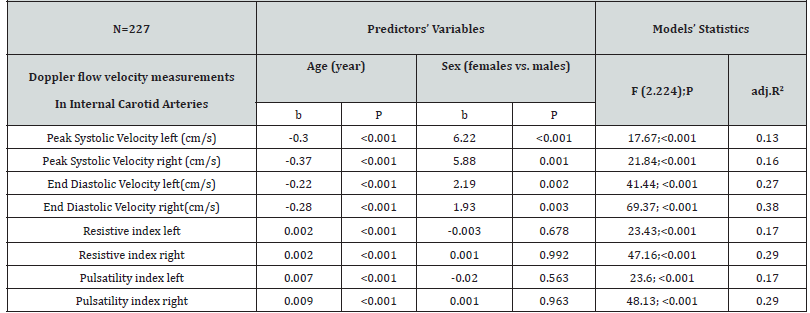
Table 2. Association of age and sex with Doppler flow velocity measurements in internal carotid arteries.
Descriptive statistic data showed that the PSV and EDV in the ICA and VA were on average higher in women than in men, due to the significant increase in velocities in women up to 60 years of age. After the menopausal years the blood flow velocities were equalized in both genders (Tables 4 & 5).
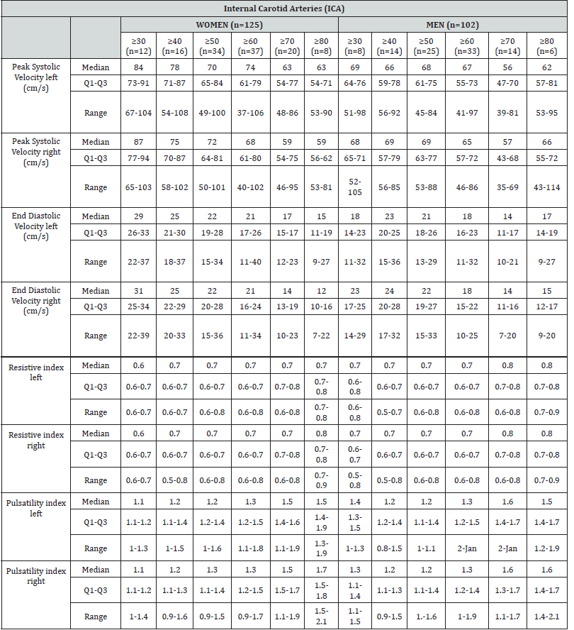
Table 4. Distribution of Doppler flow velocity measurements in internal carotid arteries in women and men in 10-year age groups.
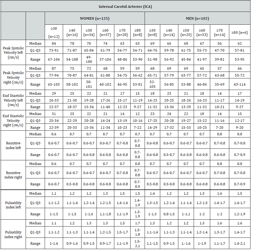
Table 5. Distribution of Doppler flow velocity measurements in vertebral arteries in women and men in 10-year age groups..
The PSV and, more pronounced, EDV in the ICA decreased with age in women. The same parameters for men showed a declining trend in the EDV, and no significant age- dependent velocity decrease in PSV, (Tables 4 & 5).
Data for the vertebral arteries showed a slight tendency to decreased flow velocities with age in the women, with the most significant decrease in EDV VA on the left side. Similar to the carotid parameters, data for the men revealed a declining trend in the EDV, with no significant age-dependent velocity decrease in PSV (Table 5).
An increase of vertebral artery diameter was found in the left vertebral artery in women in the age group ≥80. It is worth noting that this age group had a smaller number of respondents compared to the other groups.
Discussion
Knowledge on the range of velocity measurements is vital since diagnostic decisions are based on those values [15]. Data on the PSV threshold and presence of plaque represent the greatest significant factor for vascular stenosis diseases [16]. However, it is worth mentioning that normally, the brain receives a continuous arterial blood flow supply during systole and diastole due to the low resistance in the cerebral vasculature. An arterial obstruction increases vascular resistance, and the end-diastolic flow becomes affected first because it is delivered at a lower arterial blood pressure than the peak systole [17]. Low EDV seems to be associated with an increased risk of stroke and significantly increases the prediction of cardiovascular events [18,19]. The present study was designed to evaluate the normal distribution of velocity values and pulsatility and resistance parameters not only for the PSV, but also for EDV, RI, and PI in the ICA and VA, as well as the diameter of VA, as determined with carotid duplex sonography in a large population of healthy subjects. Therefore, in this study we sought to investigate the importance of comprehensive carotid Doppler measurements obtained in addition to PSV ICA and plaque measurements.
Arterial stiffening that normally occurs with age can explain the increasing flow fluctuations in cerebral blood vessels [20]. It is a proven fact that age correlates more highly with arterial stiffness than any other investigated risk factor [21]. Several studies revealed that women, in the period from adolescence to menopause, have lower arterial stiffness than age-matched men [22], but that gender differences in arterial stiffening decrease after menopause [23]. The results of our study confirmed such findings and showed that until the postmenopausal period the flow velocities (PSV and EDV) in the ICA are higher in women than in men of the same age, and that after menopause these values equalize. Moreover, according to some investigations, arteries in postmenopausal women seem to be stiffer than those of age-matched men [24]. Carotid dilation with normal aging has already been reported [25]. Hansen et al. [26] proved that the diameter of the carotid artery is significantly larger in men than in women from the age of 25, with a substantial increase until the age of 40–45 years, and an increase in older women. Tomoto et al. [27] proved that carotid arterial distensibility significantly correlated with a smaller lumen diameter and greater gradient of blood flow velocity during a cardiac cycle. In the study by Scheel P et al. [28] the ICA luminal diameters were significantly smaller in women than in men, while there was no gender difference in the diameters of the VA. The shortcoming of our study is that it did not provide information of ICA diameter.
Our results showed that in all age groups mean PSV and EDV in the ICA were on average somewhat higher in the females than in the males, which is consistent with some earlier results [8,29]. This has been attributed to the volume, which is equal to the vessel diameter multiplied by the blood flow velocity. Scheel P et al. [28] showed that, with the exception of a higher mean ICA flow velocity in women, there were no significant gender differences in any flow velocities or waveform parameters in the extracerebral vessels.
According to the investigation of carotid arteries by Eicke et al. [30], a minor but significant decrease of flow volume was found with age. Our results revealed an age-dependent and statistically significant decrease of flow velocities in the ICA. This is similar to the results published earlier [28]. It has been confirmed that diastolic carotid flow showed a significant negative correlation with the stiffness of the carotid arteries [31].
Our results showed the greatest negative correlation with age in the EDV ICA on both sides. When we separated the data for women and men, the value of PSV decreased with age in the women, while in the men there was no significant decrease; however, EDV decreased in both the men and women.
Studies of the influence of age on flow rates through the vertebral arteries have shown contradictory results. Some authors [32,33] published the results of investigations confirming that the flow rate through the vertebral arteries decreases with age, while other studies [6,34-36] did not confirm any significant differences in the diameters and flow velocity of vertebral arteries with aging. Our results showed a statistically significant age-dependent decrease of velocities in the VA, except for PSV VA on the right and the luminal diameter of the left VA. Some studies investigating vertebral artery velocities did not reveal a correlation of age and volume flow in the vertebral system [34]. A possible explanation for this phenomenon is that the brainstem is not affected by the aging process to the same extent due to the important functions it performs. The results of our study on vertebral arteries showed a slight tendency to decreased flow velocities (PSV and EDV) with age in women. Similar to the carotid parameters, data for the men revealed a declining trend in the EDV, with no significant agedependent velocity decrease in the PSV. Also, similar to the changes seen in the carotid arteries, our results suggested an equalization of flow rates between postmenopausal women and men of the same age.
Except for a very slightly higher mean PSV of the left VA in comparison with the right VA in the men, there were no significant side-to-side differences in the flow velocities and waveform parameters in the paired extracranial vessels.
Lovrencic et al. [36] showed that the mean vertebral diameter in the females was smaller and the mean blood velocity was higher than in the males, whereas the left vertebral artery was wider in both genders. Some other authors [37,38] also found that the right VA was significantly smaller. Since in some rare cases there is a dominance of the vertebral artery on the right side, our results taken together showed that the vertebral vessel diameters were wider on the left side in both genders. Scheelet al. [28] concluded that asymmetry of the luminal diameter in the vertebral arteries seems to be more the rule than the exception.
The alternative choices of Doppler speed measurements are the Doppler indices. Indices are proportions of speed measurements and thus are separate from the Doppler angle. RI and PI represent important hemodynamic parameters that have been used in the prognosis of cardiovascular complications [9]. In the renal arteries age, vascular risk factors, and clinically demonstrated vascular diseases have been proven to be associated with an increase in RI [39]. Frauchiger et al. [40] showed a positive correlation between carotid RI and the IMT as a maker of degree of atherosclerosis. This relationship could indicate that the RI ICA could be used to assess atherosclerosis. The study by Scheel et al. [28] showed a statistically significant increase in the RI in the ICA and VA with increasing age. Our results showed a slight trend of increased RI with age. Nevertheless, the RI and the range of vascular artery perfusion are inversely related. Hence, when resistance or RI increases, the perfusion will decrease.
Findings from previous studies support the hypothesis that age and arterial stiffening result in an increased carotid pulsatile flow. Mitchell et al. hypothesized that disproportionate stiffening of the proximal aorta as compared with the carotid arteries reduces wave reflection at this important interface and thereby facilitates transmission of excessive pulsatile energy into th cerebral microcirculation, leading to microvascular damage and impaired function. O’Rourke et al. [41] suggested that PI reflects the transmission of pulsatile energy into the cerebral microcirculation and highly pulsatile flow may cause brain microbleeds and lacunar infarcts. Our results showed a moderate trend of increasing PI with age. However, it is important to emphasize that the form of arterial pulse is determined by both the pattern of ventricular contraction and properties of the arterial system and arterioles [42].
Moreover, we must take into consideration that, in addition to age, blood flow velocity is influenced by vessel function, vessel size and elasticity, myocardial contractility, and cardiac output, and these parameters may vary considerably from one individual to another [43]. Like our study, studies by other authors have revealed that ranges given for the different parameters show a high interindividual variability, which results in a wide range of normal values [25,34]
In terms of study limitations, we must emphasize that we have not been able to fully control risk factor for cerebrovascular disease (eg. “well-controlled arterial hypertension”) and/or some exclusion criteria (eg. pregnancy, malignancies), and the observational nature of this design leaves the possibility of residual confounding
We can conclude that the Vascular Laboratory hospital protocols should require measurement and recording of all relevant information related to extracranial artery color Doppler assessment. Based on the results of our research and some previous studies normal range rather than rigid cutoff values for hemodynamics parameters in the extracranial part of the ICA and VA adjusted for age and sex remain to be further investigated and defined.
Acknowledgment
The authors wish to thank Aleksandra Zmegac Horvat for language editing the manuscript.
Conflict of Interest
No potential conflict of interest relevant to this article has been reported.
References
- Mattle HP Kent KC, Edelman RR, Atkinson DJ, Skillman JJ (1991) Evaluation of the extracranial carotid arteries. J Vasc Surg 13(6): 838-844.
- Jahromi AS, Cinà CS, Liu Y, Clase CM (2005) Sensitivity and specificity of color duplex ultrasound measurement in the estimation of internal carotid artery stenosis: A systematic review and meta-analysis. J Vasc Surg 41(6): 962-972.
- Pourcelot L (1975) Applications cliniques de l′examen Doppler transcutan. Velocim Doppler 1975: 213-240.
- Wielicka M, Neubauer-Geryk J, Kozera G, Bieniaszewski L (2020) Clinical application of pulsatility index. Med Res J 5(3): 201-210.
- Hirata K, Yaginuma T, O’Rourke MF, Kawakami M (2006) Age-related changes in carotid artery flow and pressure pulses: Possible implications for cerebral microvascular disease. Stroke 37(10): 2552-2556.
- Nemati M, Bavil AS, Taheri N (2009) Comparison of normal values of Duplex indices of vertebral arteries in young and elderly adults. Cardiovasc Ultrasound 7: 2.
- Albayrak R, Degirmenci B, Acar M, Haktanir A, Colbay M, et al. (2007) Doppler sonography evaluation of flow velocity and volume of the extracranial internal carotid and vertebral arteries in healthy adults. J Clin Ultrasound 35(1): 27-33.
- Bello AB, Bello Tom Aremu AA, Adetiloye VA (2016) Normal cerebral blood flow volume in healthy Nigerian adults. Br J Med Med Res 17(2): 1-11.
- Staub D, Meyerhans A, Bundi B, Schmid HP, Frauchiger B (2006) Prediction of cardiovascular morbidity and mortality: Comparison of the internal carotid artery resistive index with the common carotid artery intima-media thickness. Stroke 37(3): 800-805.
- Kumar LA, Nagarajan C (2015) Common carotid artery intima media thickness segmentation survey. World Acad Sci Eng Technol. Int J Med Heal Biomed Bio eng Pharm Eng 8(9): 689-693.
- Unal B, Bagcier S, Simsir I, Bilgili Y, Kara S (2004) Evaluation of differences between observers and automatic-manual measurements in calculation of Doppler parameters. J Ultrasound Med 23(8): 1041-1048.
- Nelson TR, Pretorius DH (1988) The Doppler signal: Where does it come from and what does it mean? American Journal of Roentgenology 151(3): 439-447.
- Michel E, Zernikow B (1998) Gosling’s doppler pulsatility index revisited. Ultrasound Med Biol 24(4): 597-599.
- Almuhanna K, Zhao L, Kowalewski G, Beach KW, Lal BK, et al. (2012) Investigation of cerebral hemodynamics and collateralization in asymptomatic carotid stenoses. Annu Int Conf IEEE Eng Med Biol Soc 2012: 5618-5621.
- Kalvach P, Gregová D, Škoda O, Peisker T, Tůmová R, et al. (2007) Cerebral blood supply with aging: Normal, stenotic and recanalized. J Neurol Sci 257(1-2): 143-148.
- Grant EG, Benson CB, Moneta GL, Alexandrov AV, Baker JD, et al. (2003) Carotid Artery Stenosis: Gray-Scale and Doppler US Diagnosis - Society of Radiologists in Ultrasound Consensus Conference. Radiology 229(2): 340-346.
- Alexandrov AV, Tsivgoulis G, Rubiera M, Vadikolias K, Stamboulis E, et al. (2010) End-diastolic velocity increase predicts recanalization and neurological improvement in patients with ischemic stroke with proximal arterial occlusions receiving reperfusion therapies. Stroke 41(5): 948-952.
- Chuang SY, Bai CH, Chen JR, Yeh WT, Chen HJ, et al. (2011) Common Carotid End-Diastolic Velocity and Intima-Media Thickness Jointly Predict Ischemic Stroke in Taiwan. Stroke 42(5): 1338-1344.
- Chuang SY, Bai CH, Cheng HM, Chen JR, Yeh WT, et al. (2016) Common carotid artery end-diastolic velocity is independently associated with future cardiovascular events. Eur J Prev Cardiol 23(2): 116-124.
- Smulyart H, Asmar RG, Rudnicki A, London GM, Safar ME (2001) Comparative effects of aging in men and women on the properties of the arterial tree. J Am Coll Cardiol 37(5): 1374-1380.
- El Feghali R, Topouchian J, Pannier B, Asmar R (2007) Ageing and blood pressure modulate the relationship between metabolic syndrome and aortic stiffness in never-treated essential hypertensive patients. A comparative study. Diabetes Metab 33(3): 183-188.
- Ahimastos AA, Formosa M, Dart AM, Kingwell BA (2003) Gender Differences in Large Artery Stiffness Pre- and Post Puberty. J Clin Endocrinol Metab 88(11): 5375-5380.
- Weng C, Yuan H, Yang K, Tang X, Huang Z, et al. (2013) Gender-specific association between the metabolic syndrome and arterial stiffness in 8,300 subjects. Am J Med Sci 346(4): 289-294.
- Noon JP, Trischuk TC, Gaucher SA, Galante S, Scott RL (2008) The effect of age and gender on arterial stiffness in healthy Caucasian Canadians. J Clin Nurs 17(7): 2311-2317.
- Schoning M, Walter J, Scheel P (1994) Estimation of cerebral blood flow through color duplex sonography of the carotid and vertebral arteries in healthy adults. Stroke 25(1): 17-22.
- Hansen F, Mangell P, Sonesson B, Länne T (1995) Diameter and compliance in the human common carotid artery - variations with age and sex. Ultrasound Med Biol 21(1): 1-9.
- Tomoto T, Maeda S, Sugawara J (2017) Influence of blood flow velocity on arterial distensibility of carotid artery in healthy men. J Physiol Sci 67(1): 191-196.
- Scheel P, Ruge C, Schöning M (2000) Flow velocity and flow volume measurements in the extracranial carotid and vertebral arteries in healthy adults: Reference data and the effects of age. Ultrasound Med Biol 26(8): 1261-1266.
- Yazici B, Erdogmus B, Tugay A (2005) Cerebral blood flow measurement of extracranial carotid and vertebral Doppler ultrasonography in healthy adults. DiagnInterv Radiol 11(4): 195-198.
- Weskott HP, Holsing K (1997) US-based evaluation of hemodynamic parameters in the common carotid artery: a nomogram trial. Radiology 205(2): 353-359.
- Jiang YN, Kohara K, Hiwada K (1998) Alteration of carotid circulation in essential hypertensive patients with left ventricular hypertrophy. J Hum Hypertens 12(3): 173-179.
- Müller M, Schimrigk K (1994) A comparative assessment of cerebral haemodynamics in the basilar artery and carotid territory by transcranial Doppler sonography in normal subjects. Ultrasound Med Biol 20(8): 677-687.
- Martin PJ, Evans DH, Naylor AR (1994) Transcranial color-coded sonography of the basal cerebral circulation; reference data from 115 volunteers. Stroke 25(2): 390-396.
- Seidel E, Eicke BM, Tettenborn B, Krummenauer F (1999) Reference values for vertebral artery flow volume by duplex sonography in young and elderly adults. Stroke 30(12): 2692-2696.
- Trattnig S, Hübsch P, Schuster H, Pölzleitner D (1990) Color-coded doppler imaging of normal vertebral arteries. Stroke 21(8): 1222-1225.
- Lovrencic-Huzjan A, Demarin V, Bosnar M, Vukovic V, Podobnik-Sarkanji S (1999) Color Doppler Flow Imaging (CDFI) of the vertebral arteries - The normal appearance, normal values and the proposal for the standards. Coll Antropol 23(1): 175-181.
- Simon H, Niederkorn K, Horner S, Duft M, Schrockenfuchs M. Einfluss der Halsdrehung auf das vertebrobasiläre System. (1994) Eintranskraniell-dopplersonographischerBeitragzurPhysiologie. HNO. (42):614–8.
- Schöning M, Hartig B (1998) The development of hemodynamics in the extracranial carotid and vertebral arteries. Ultrasound Med Biol 24(5): 655-662.
- Ishimura E, Nishizawa Y, Kawagishi T, Okuno Y, Kogawa K, et al. (1997) Intrarenal hemodynamic abnormalities in diabetic nephropathy measured by duplex Doppler sonography. Kidney int 51(6): 1920-1927.
- Frauchiger B, Peter Schmid H, Roedel C, Moosmann P, Staub D (2001) Comparison of carotid arterial resistive indices with intima-media thickness as sonographic markers of atherosclerosis. Stroke 32(4): 836-841.
- O’Rourke MF, Safar ME (2005) Relationship between aortic stiffening and microvascular disease in brain and kidney: Cause and logic of therapy. Hypertension 46(1): 200-204.
- Vlachopoulos C, O’Rourke M, Nichols WW (2011) McDonald’s Blood Flow in Arteries. CRC Press.
- Blackshear WM, Phillips DJ, Chikos PM, Harley JD, Thiele BL, et al. (1980) Carotid artery velocity patterns in normal and stenotic vessels. Stroke 11(1): 67-71.



 We use cookies to ensure you get the best experience on our website.